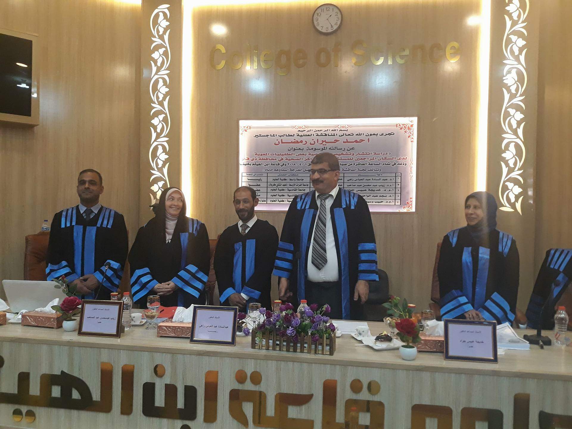
على قاعة أبن الهيثم في كلية العلوم بجامعة القادسية جرت مناقشة الدراسة المُعَدة من قبل الطالب احمد حيران رمضان تحت عنوان التحري الوراثي الجزيئي عن بعض الطفيليات المعوية لمراجعي المستشفيات والمراكز الصحية في محافظة ذي قار دراسة بأشراف أ.د نجم عبد الواحد عبد الخضر(رحمه الله) وبعضوية ودفاع السيد ا.م.د.حبيب وسيل شبر(لوفاة المشرف) … وبينت الدراسة التي أجريت في معظم أقضية محافظة ذي قار ونواحيها من بداية تشرين الأول لعام 2016وحتى نهاية شهر تموز للعام 2017 بهدف تحديد انتشار الطفيليات المعوية بين المراجعين إلى المؤسسات الصحية في المحافظة وتشخيصها ودراسة أمراضيتها من خلال جمع 1393 عينة براز لذكور وإناث (816 و 577) علـــى التوالي، إن عدد المصابين بالطفيليات المعوية 506 مصاب من بين 1393مفحوصاً أي بنسبة إصابة بلغت 36.3% إذ كانت الاصابة للذكور286((%20.5 ونسبة الاصابة للاناث 15.7)220%).
توصلت هذه الدراسة الى تشخيص 11 نوعاً من الطفيليات المعوية 9 منها شخصت بالمجهر وهي طفيلي الاميبا الحالة للنسيج Entamoeba histolyticaبنسبة %86.3 و طفيلي الجيارديا اللامبيلية Giardia lambliaبنسبة 7.5%, طفيلي الابواغ الخبيئة Cryptosporidium parvum والمتحولة القولونيةEntamoeba coli والمشعرة البشرية Trichomonas hominis كانت %1.1,%0.3,%0.1 على التوالي.كذلك الديدان المسجلة من هذه الدراسة فهي الديدان الدبوسية Enterobius vermicularis وديدان الصــــــفر الخراطــــــــيني Ascaris lumbricoides والدودة الشريطية محرشفة الغشاءHymenolepis ssp. ودودة البقــر الشـــريطــــية Taenia saginataبالـــنســـب %5.7, 0.7% ,0.6% , 0.1% على الترتيب, اما النوعان E.dispar و E.hartmanni تم التأكد منهما بتقنية الــPCR لعدم إمكانية تمييزهما عن طفيلي E.histolyticaبالفحص ألمجهري وظهرا بالنسب %8.3,%14.5 على التوالي. بالإضافة إلى تأكيد التشخيص لعينات الاميبا الحالة للنسيج والدودة الدبوسية والصفر الخراطيني بطريقة الـ PCR.
سجل شهر تشرين الأول أعلى نسبة إصابة بلغت %55.5 وأعلى الفئات العمرية بالإصابة كانت الفئة 10-6 بنسبة %42.9كما إن اغلب أنواع الإصابات كانت هي الإصابة المفردة %97 ورافقت هذه الإصابات الأعراض المرضية كالإسهال والحمى والتقيؤ وآلام البطن وبعضها كان من دون أعراض سريرية .كما سجلت الدراسة فروقاً معنوية لبعض مكونات الدم لدى المصابين بالطفيليات المعوية كنسبة الهيموكلوبين وحجم كريات الدم المضغوط والعدد الكلي والتفريقي لخلايا الدم البيض.
كمـــــــــــــا تم اســـــــــــــــتعمال طــــــــــــــــــرائق التشخيــــــــص الجـــــــــــزيئي باستعمال PCR والشجرة الوراثية التطورية ,Phylogenetic treeوتسلسل الدنا DNA Sequencing وذلك لتأكيد نتائج الفحص ألمجهري والتمييز الدقيق لأنواع جنس Entamoeba spp. عن بعضـــــــــها اعتـــــــــمادا على الجــــــــــــــين (18S rRNA) وكذلك تم إجراء تحليل تتابع الحمض النووي وتحليل الشجرة الوراثية بحسب الطريقة Joining- Neighborللأنواع الثلاثة لغرض مقارنتها مع العزلات المختلفة باستعمال البرنامج . MEGA6 ولمقارنة نسب التشابه فيما بين سلالات هذه الدراسة مع العزلات العالمية والمسجلة ببنك الجينات العالمي NCBI ضمن قاعدة البيانات Data base تم استعمال برنامج NCBI .Blast كما سجلت العزلات لانواع الـــــــــ Entamoeba بعد إن أكدت بالفحص الجزيئي في NCBIلبنك الجينات العالمي وتم تزويدنا بالأرقام التسلسلية العالمية لعزلة كل نوع من الأنواع الثلاثة .
ومن الجدير بالذكر إن العزلة E.hartmanniلم تسجل سابقا في بنك الجينات العالمي كعزلة عراقية لذلك تم تسجيلها من خلال الدراسة الحالية وبذلك هي الأولى من نوعها سجلت في بنك الجينات العالمي.فضلا عن تأكيد التشخيص ألمجهري للنوعين Enterobius vermicularis و Ascaris lumbricoides بطريقة الفحص الجزيئي (PCR).
This study was conducted in most areas and districts of Thi Qar province from the beginning of October 2016 until the end of July 2017 in order to determine the distributed of intestinal parasites among the population to attending the health institutions in the province and diagnose and study its morbidity through collecting 1393 stool samples of male and female (816 and 577) respectively. It was found that the number of infected intestinal parasites 506 infected among 1393 subjects, The Percentage of infection it 36.3% of the rate was 286 males by 20.5% and 220 females by 15.7%.
The study identified 11 types of intestinal parasites, 9 of which were diagnosed with Entamoeba histolytica (87.3%), Giardia lamblia (7.5%), Cryptosporidium parvum , Entamoeba coli and Trichomonas hominis (1.1%, 0.3%, 0.1% ) respectively. The worms recorded from this study were the Enterobius vermicularis , Ascaris lumbricoides, Hymenolepis spp. , and Taenia saginata (5.7%, 0.7%, 0.6%, 0.1%), respectively. As for E.dispar and E.hartmanni were Confirmation of the PCR method for not being distinguishable from E. histolytica parasites by microscopic examination and by percentage (8.3%, 14.5%) respectively. Added to confirm the diagnosis of samples E. histolytica and Enterobius vermicularis and Ascaris lumbricoides to molecular diagnostic by PCR technique.
The highest rate of infection was recorded October (55.5%) and the highest age group was 6-10 years (42.9%), and most cases of infections were single infection 97%, accompanied by symptoms such as diarrhea, fever, vomiting and abdominal pain, but some of them asymptoms. The study also showed significant differences in some blood components in patients with intestinal parasites such as hemoglobin, packed cells volume , total and differential blood white blood count.
Molecular diagnostic methods using PCR, Phylogenetic tree and DNA Sequencing were used to confirm the results of microscopic examination and the precise differentiation of the Entamoeba spp. depending on (18S rRNA). DNA sequence analysis and gene analysis of the three species were performed to compare them with different species using MEGA6 and to compare the similarities between the strains of this study with the global species recorded in the NCBI database by using Blast program. The species of Entamoeba were recorded after confirming the NCBI Molecular Examination of the Global Gene Bank and provided us with global serial numbers to isolate each of the three species. It is worth noting that the isolation of E.hartmanni was not previously registered in the World Bank of Genes as an Iraqi isolation, so it was recorded through the current study and thus is the first of its kind recorded in the World Bank of Genetics. In addition to confirm the microscopic diagnosis of the types Enterobius vermicularis and Ascaris lumbricoides by polymerase chain reaction.